RETURN OF THE DEV BIO IMAGE CONTEST!

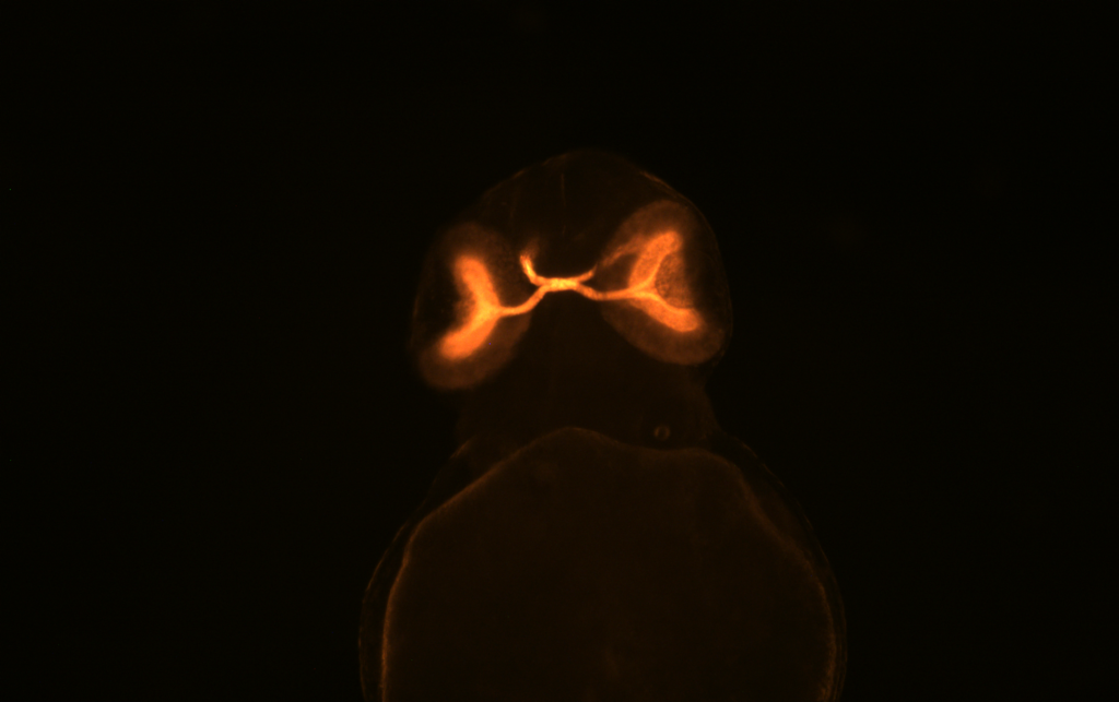
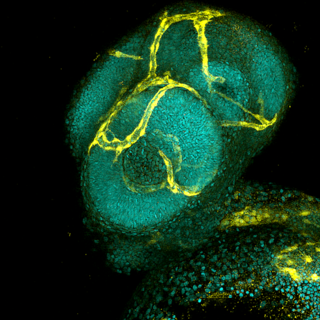
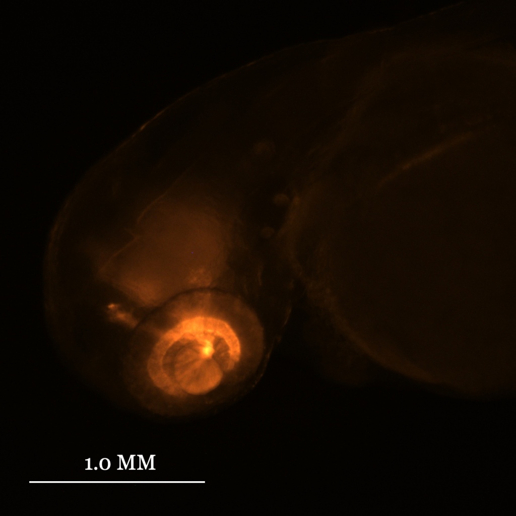
After a two-year hiatus, Developmental Biology (BIO 351L) at Reed College is back — in person and imaging lots of zebrafish embryos. This year’s image contest featured 9 entries from 7 students. Thumbnails of all the entries are below. All images were captured with light microscopy by undergraduates who investigated cell proliferation, the expression of understudied genes, neuron projections, and blood vessel formation in the developing zebrafish visual system.

And the winner is…

As part of a study to examine blood vessel development in gdf6a mutant fish, Reed College juniors, Jiahe Yue and Frank Zhuang, captured images of 3 day old wild-type (shown above) and mutant (not shown) zebrafish embryos expressing kdrl:GFP to highlight blood vessel formation (yellow). They counterstained nuclei with DAPI (cyan). Image is a maximum intensity projection using a 25x 1.1 NA objective on a Nikon A1+ laser scanning confocal microscope in the Cerveny Lab at Reed College.

Leave a comment